38 external structure of the heart with labels
Dapagliflozin - Wikipedia Medical uses. Dapagliflozin is used along with diet, exercise and usually with other glucose lowering medications, to improve glycaemic control in adults with type 2 diabetes and to reduce the risk of hospitalisation for heart failure among adults with type 2 diabetes and known cardiovascular disease or other cardiovascular risk factors (including high blood pressure, high cholesterol and ... Label the heart — Science Learning Hub Label the heart Interactive Add to collection In this interactive, you can label parts of the human heart. Drag and drop the text labels onto the boxes next to the diagram. Selecting or hovering over a box will highlight each area in the diagram. pulmonary vein semilunar valve right ventricle right atrium vena cava left atrium pulmonary artery
Diagram of the human heart royalty-free images - Shutterstock 14,791 diagram of the human heart stock photos, vectors, and illustrations are available royalty-free. See diagram of the human heart stock video clips Image type Orientation Color People Artists Sort by Popular Anatomy Healthcare and Medical Icons and Graphics Diseases, Viruses, and Disorders heart medicine organ hemodynamics circulatory system

External structure of the heart with labels
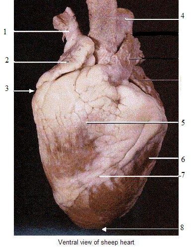
Heart Anatomy: Heart Dissection - University of Washington The letters indicated in the text refer to the labels on the picture. The anterior surface of the heart is characterized by the presence of the large arteries leaving the base of the heart, the pulmonary trunk (H) and the aorta (G). The pulmonary trunk is the vessel that divides to give rise to the two pulmonary arteries going to each lung. Label the Heart - The Biology Corner Shows a picture of a heart with letters and blanks for practice with labeling the parts of the heart and tracing the flow of blood within the heart. Heart Anatomy: size, location, coverings and layers : Anatomy & Physiology The heart wall is composed of three layers: the epicardium, myocardium, and endocardium. Location of the heart in the mediastinum. The superficial epicardium is the visceral layer of the serous pericardium. The middle layer is the myocardium and is composed mainly of cardiac muscle and forms the bulk of the heart.
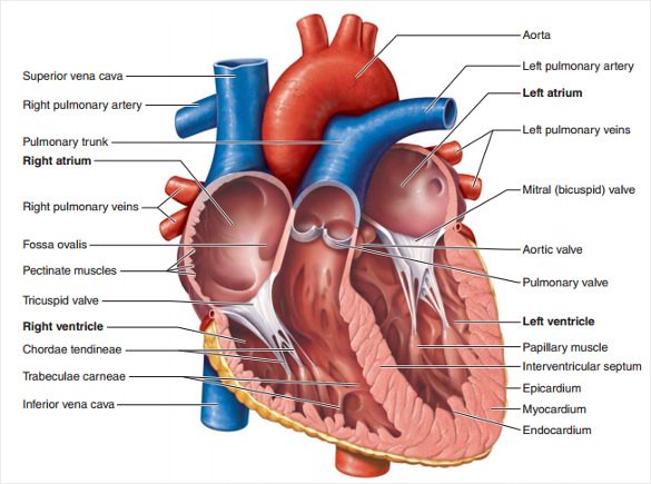
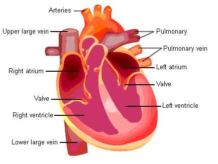
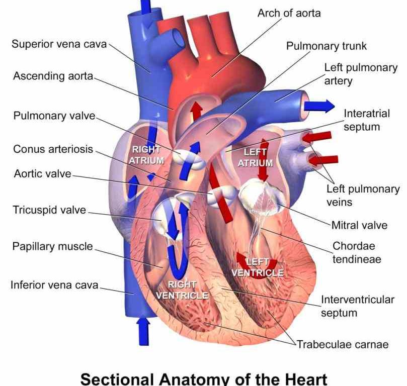
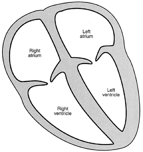
External structure of the heart with labels. Labelling the heart — Science Learning Hub Blood transports oxygen and nutrients to the body. It is also involved in the removal of metabolic wastes. In this interactive, you can label parts of the human heart. Drag and drop the text labels onto the boxes next to the diagram. Selecting or hovering over a box will highlight each area in the diagram. Heart Anatomy | Anatomy and Physiology | | Course Hero The cardiovascular system is a closed system if the heart and blood vessels. The heart pumps blood through a closed system of blood vessels. Blood vessels allow blood to circulate to all parts of the body. Arteries usually colored red because oxygen rich, carry blood away from the heart to capillaries within the tissues. Human Heart Diagram Labeled | Science Trends The endocardium is the inner portion of the outer wall, and the endocardium is what contacts the blood in the heart. The heart's atrioventricular valves are structures that join the atria and ventricles of the heart together. This group of valves is comprised of the tricuspid valve and the mitral valve. A Labeled Diagram of the Human Heart You Really Need to See The human heart, comprises four chambers: right atrium, left atrium, right ventricle and left ventricle. The two upper chambers are called the left and the right atria, and the two lower chambers are known as the left and the right ventricles. The two atria and ventricles are separated from each other by a muscle wall called 'septum'.
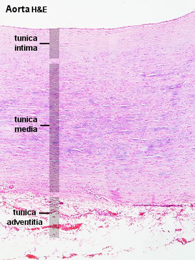
Layers of the heart: Epicardium, myocardium, endocardium - Kenhub The myocardium is functionally the main constituent of the heart and the thickest layer of all three heart layers. It is a muscle layer that enables heart contractions. Histologically, the myocardium is comprised of cardiomyocytes.Cardiomyocytes have a single nucleus in the center of the cell, which helps to distinguish them from skeletal muscle cells that have multiple nuclei dispersed in the ... Heart Diagram with Labels and Detailed Explanation - BYJUS Diagram of Heart. The human heart is the most crucial organ of the human body. It pumps blood from the heart to different parts of the body and back to the heart. The most common heart attack symptoms or warning signs are chest pain, breathlessness, nausea, sweating etc. The diagram of heart is beneficial for Class 10 and 12 and is frequently ... Chapter 19: The Heart Flashcards | Quizlet Heart External Anatomy- Anterior Chambers. See image. ... Place the labels in order denoting the flow of blood through the pulmonary circuit beginning with the right atrium and ending in the ... Impulse conduction through the cardiac conduction system is slowest through which structure, thereby allowing a pause between atrial contraction and ... How to Use Microsoft Access (with Pictures) - wikiHow 18.3.2022 · Determine the best structure for your data. If you are creating a blank database, you’ll want to think about the best way to organize your data, and add the appropriate structure. There are several ways that you can format and interact with your data in Access: Tables – This is the main way that data is stored in your database.
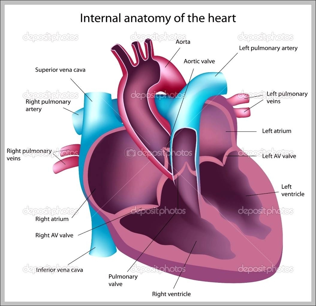
Correctly Label The Following External Anatomy Of The Anterior Heart ... The external anatomy of the human heart consists of the four chambers that form the apex of the heart. Each chamber has an apex that corresponds to a box. There are two boxes on each side of the heart: the atria and the ventricles. The left atrium is a branching organ. The pulmonary trunk contains the aorta and pulmonary veins. Structure of the Heart | SEER Training - National Cancer Institute The human heart is a four-chambered muscular organ, shaped and sized roughly like a man's closed fist with two-thirds of the mass to the left of midline. The heart is enclosed in a pericardial sac that is lined with the parietal layers of a serous membrane. The visceral layer of the serous membrane forms the epicardium. Layers of the Heart Wall Heart Anatomy: Labeled Diagram, Structures, Blood Flow ... - EZmed There are 4 chambers, labeled 1-4 on the diagram below. To help simplify things, we can convert the heart into a square. We will then divide that square into 4 different boxes which will represent the 4 chambers of the heart. The boxes are numbered to correlate with the labeled chambers on the cartoon diagram. View fullsize Human Heart - Anatomy, Functions and Facts about Heart - BYJUS The external structure of the heart has many blood vessels that form a network, with other major vessels emerging from within the structure. The blood vessels typically comprise the following: Veins supply deoxygenated blood to the heart via inferior and superior vena cava, and it eventually drains into the right atrium.
Things cardiologists never tell you: KNOW YOUR HEART. Structure of the heart and blood vessels ...
The Anatomy of the Heart, Its Structures, and Functions - ThoughtCo The heart is the organ that helps supply blood and oxygen to all parts of the body. It is divided by a partition (or septum) into two halves. The halves are, in turn, divided into four chambers. The heart is situated within the chest cavity and surrounded by a fluid-filled sac called the pericardium. This amazing muscle produces electrical ...
13+ Heart Diagram Templates – Sample, Example, Format Download | Free & Premium Templates
External anterior heart labeling Quiz - purposegames.com This is an online quiz called External anterior heart labeling. There is a printable worksheet available for download here so you can take the quiz with pen and paper. Your Skills & Rank. Total Points. 0. ... Internal Anatomy of the Kidney 4p Image Quiz. Arteries of the abdomen view 2 6p Image Quiz. Arteries of the abdomen view 3 5p Image Quiz.
Heart Model Contiunued - ProProfs Quiz
What is a Medical Device Technical File and How to Structure It? 16.5.2022 · At the heart of the approval process for your medical devices is the medical device technical documentation. This file is not only required for approval to sell your medical devices in the EU, but they are also subject to regular inspection by the US FDA and ISO 13485:2016 audits.
Know the Structures and Functions about Your Heart | New Health Advisor
Heart - External Features - Anatomy QA Location of heart: Heart lies in the middle mediastinum. 1/3rd of the heart lies to the right and 2/3rd to the left of the midline. It lies opposite to T5 - T8 vertebrae in supine position & T6 - T9 vertebrae in erect position. Dimensions of heart: Base to apex-12cm; Transversely- 8-9cm; Anteroposteriorly- 6cm.
called myocardium science External Structure Of Human Heart Anatomy structure of human heart ...
NASA - NASA Facilities By Frank Seitzen “Houston, Tranquility Base here. The Eagle has landed.” Bold goals - President George W. Bush announced new space exploration goals for the agency at NASA Headquarters on Jan. 14, 2004. Photo credit: NASA/Bill Ingalls. When a city’s name is one of the first words spoken on the surface of another world, the link between that city and NASA through its …

Lung Structure | BioNinja
Heart anatomy: Structure, valves, coronary vessels | Kenhub The heart is shaped as a quadrangular pyramid, and orientated as if the pyramid has fallen onto one of its sides so that its base faces the posterior thoracic wall, and its apex is pointed toward the anterior thoracic wall.

Biology A&P Lab: Study guide for quiz on Feb.25th
How Strategy Shapes Structure - Harvard Business Review See Industrial Market Structure and Economic Performance, F. M. Sherer (Chicago: Rand McNally, 1970). 2. See Blue Ocean Strategy , W. Chan Kim and Renée Mauborgne (Harvard Business Press, 2005).
How would you label the structures (both external and internal) of a dissected pig's heart? - Quora
Heart Labeling Quiz: How Much You Know About Heart Labeling? Here is a Heart labeling quiz for you. The human heart is a vital organ for every human. The more healthy your heart is, the longer the chances you have of surviving, so you better take care of it. Take the following quiz to know how much you know about your heart. Questions and Answers. 1.
ANAT2241 Cardiovascular System - Embryology
Chapter 22 Heart Flashcards | Quizlet Label the coronary arteries in an anterior view of the heart. Label the order that blood flows through in the heart, using the arrows as guides. Label the components of the heart wall. Label the components of the heart as seen from a posterior view. Label the major coronary veins. Label the components of the conduction system.

Heart Structure
The Tenors - Wikipedia The Tenors (formerly known as The Canadian Tenors) are a vocal group consisting of Victor Micallef, Clifton Murray, Alberto Urso, and Mark Masri.They perform operatic pop music that is a mixture of classical and pop, featuring songs such as "The Prayer", Panis angelicus, and Leonard Cohen's Hallelujah.. Originating from Canada, Micallef and Masri from Toronto, Urso from …

Knowledge Class: The Heart
Structure and Function of the Heart - News-Medical.net Structure of the heart. The heart wall is composed of three layers, including the outer epicardium (thin layer), middle myocardium (thick layer), and innermost endocardium (thin layer). The ...

CLASS BLOG: BIO 202 Heart Anatomy Worksheet
How to Analyze Sentences (with Pictures) - wikiHow 21.1.2022 · Use labels according to how much of the sentence you’re analyzing and the components that make that part up. If you’re analyzing a complete sentence or a clause, you’re looking for a subject and a predicate. Your subject will be a noun phrase, while your predicate will be a verb phrase.
Structure and Function of the Normal Heart | Thoracic Key
Solved Correctly label the following external anatomy of the - Chegg Ans : 1: Ascending aorta. Ascending aorta is the largest artery, carrying oxygenated blood from left ventricles to the body part. It arises from the left ventricles. 2: Ligamentum arteriosum It is a vestigi …. View the full answer. Transcribed image text: Correctly label the following external anatomy of the anterior heart.

38 Label The Following Diagram Of The Heart - Labels 2021
Lesson | The Heart - External Structure | Encounter Edu In this lesson students begin their exploration of the circulatory system, labelling a diagram of the external structures and identifying arteries and veins. They will go on to explain where blood enters and leaves the heart. Learning outcomes

what is heart!?what is it function? and structure - Brainly.in
VP Online - Online Drawing Tool - Visual Paradigm VP Online is your all-in-one online drawing solution. Create professional flowcharts, UML diagrams, BPMN, ArchiMate, ER Diagrams, DFD, SWOT, Venn, org charts and mind map. Works cross-platforms: Mac, Windows, Linux. Sign-up for a FREE account today!
37 Label The Anatomy Of The Heart - Labels 2021
2. External features of the heart - SlideShare 2. THE HEART • The heart is a hollow muscular organ that is pyramidal in shape • It lies within the pericardium in the middle mediastinum • It is connected at its base to the great blood vessels. 3. General features of the heart • The heart has; • an apex and base • 2 surfaces; • Sternocostal surface • Diaphragmatic surface ...

Free Blank Heart Diagram, Download Free Blank Heart Diagram png images, Free ClipArts on Clipart ...
Solved Art-Labeling Activity: Overview of the external - Chegg art-labeling activity: overview of the external anatomy of the heart anterior view res great cardiac vein aortic arch right coronary artery left coronary artery left pulmonary veins ascending aorta left pulmonary artery anterior interventricular artery superior vena cava pulmonary trunk auricle of left atrium circumflex artery auricle of right …

fantasticvoyagewiki / Heart
Internal Structure of the Heart | Contemporary Health Issues It is marked by the presence of four openings that allow blood to move from the atria into the ventricles and from the ventricles into the pulmonary trunk and aorta. Located in each of these openings between the atria and ventricles is a valve, a specialized structure that ensures one-way flow of blood.
Post a Comment for "38 external structure of the heart with labels"