39 cell diagram and labels
Plant Cell: Diagram, Types and Functions - Embibe Exams Q.2. How to make a model of a plant cell diagram step by step procedure? Ans: The plant cell diagram can be checked above and on a similar pattern the diagram can be created. Q.3. Why do plant cells possess large-sized vacuoles? Ans: Vacuole functions in the storage of substances, maintenance of osmolarity and sustaining turgor pressure. Q.4. Cell Diagram | Free Cell Diagram Templates - Edrawsoft A free customizable cells diagram template is provided to download and print. Quickly get a head-start when creating your own cell diagram. Here is a simple cell diagram example created by Science Diagram Maker Download Template: Get EdrawMax Now! Free Download Popular Latest Flowchart Process Flowchart Workflow BPMN Cross-Functional Flowchart
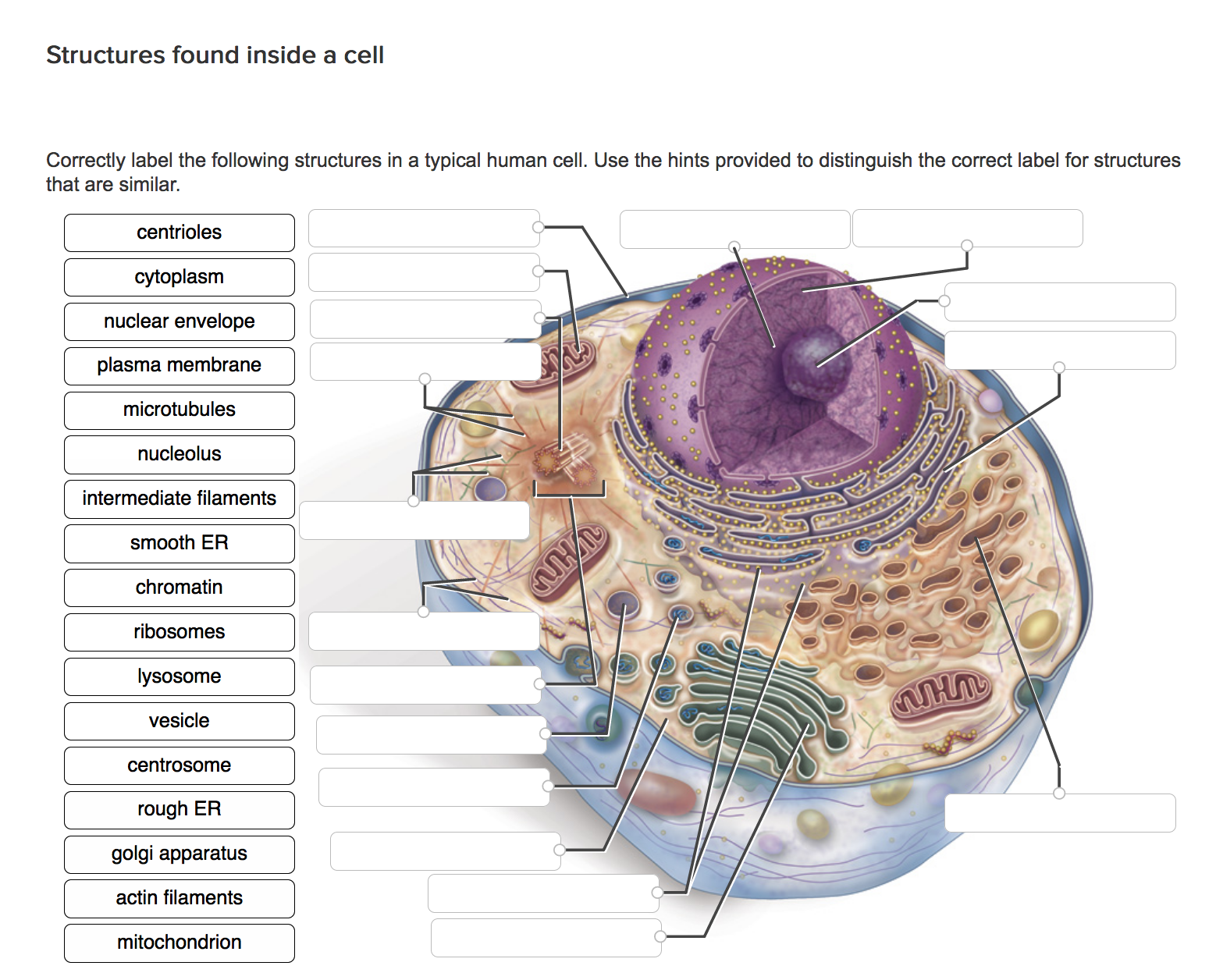
PDF Human Cell Diagram, Parts, Pictures, Structure and Functions One of the few cells in the human body that lacks almost all organelles are the red blood cells. The main organelles are as follows : cell membrane endoplasmic reticulum Golgi apparatus lysosomes mitochondria nucleus perioxisomes microfilaments and microtubules 2
Cell diagram and labels
A Labeled Diagram of the Animal Cell and its Organelles A Labeled Diagram of the Animal Cell and its Organelles There are two types of cells - Prokaryotic and Eucaryotic. Eukaryotic cells are larger, more complex, and have evolved more recently than prokaryotes. Where, prokaryotes are just bacteria and archaea, eukaryotes are literally everything else. Converting Diagrams - The Biology Corner Open Google Draw and import the diagram. Then use "insert" to create text boxes where students can fill in the labels. Don't forget when assigning this to students on Google classroom to make a copy for each student. You can leave documents in an uneditable form and students can use an addon like "Kami" to annotate the document. Label the cell - Teaching resources - Wordwall by Mbauer. Correctly Label the Bacteria (Prokaryotic) Cell Labelled diagram. by Bronwyn12. Label Plant and Animal Cell Labelled diagram. by Catherine34. Plant Cell - Label Organelles Labelled diagram. by Azimmer. Animal Cell Label Labelled diagram. by Taraabbott.
Cell diagram and labels. 2,119 Red blood cell diagram Images, Stock Photos & Vectors - Shutterstock Find Red blood cell diagram stock images in HD and millions of other royalty-free stock photos, illustrations and vectors in the Shutterstock collection. Thousands of new, high-quality pictures added every day. cell membrane diagram with labels cell membrane diagram with labels Structure protein cell each function animal gene labels label following masteringbiology synthesis diagram steps processes locations involved identify appropriate process. Cell parts label animal plant membrane worksheet cells labeled structure labels functions worksheets diagrams visit. Cell diagram with labels - Graph Diagram This human anatomy diagram with labels depicts and explains the details and or parts of the Cell Diagram With Labels.Human anatomy diagrams and charts show internal organs, body systems, cells, conditions, sickness and symptoms information and/or tips to ensure one lives in good health. Free Cell Diagram Software with Free Templates - EdrawMax - Edrawsoft An animal cell diagram describes a cell structure enclosed by a plasma member, and it has a nucleus with a membrane and organelles. Neuron Diagram A neuron diagram describes the three parts of a Neuron: dendrites, an axon, a cell body, or soma. Cell Membrane Diagram
A Labeled Diagram of the Plant Cell and Functions of its Organelles ... The cell membrane is a thin layer made up of proteins, lipids, and fats. It forms a protective wall around the organelles contained within the cell. It is selectively permeable and thus, regulates the transportation of materials needed for the survival of the organelles of the cell. Function: Protects the cell from its surroundings. Learn the parts of a cell with diagrams and cell quizzes Cell diagram unlabeled It's time to label the cell yourself! As you fill in the cell structure worksheet, remember the functions of each part of the cell that you learned in the video. Doing this will help you to remember where each part is located. Click the links below to download the labeled and unlabeled eukaryotic cell diagrams. A Well-labelled Diagram Of Animal Cell With Explanation - BYJUS The animal cell diagram is widely asked in Class 10 and 12 examinations and is beneficial to understand the structure and functions of an animal. A brief explanation of the different parts of an animal cell along with a well-labelled diagram is mentioned below for reference. Also Read Different between Plant Cell and Animal Cell Animal Cell Diagram with Label and Explanation: Cell ... - Collegedunia Diagram of Animal Cell Below is the diagram of the animal cell which shows the organelles present in it. The cell is covered with cytoplasm which consists of cell organelles in it. The nucleus is covered with a rough Endoplasmic Reticulum and other organelles each designed for a specific purpose.
Cell: Structure and Functions (With Diagram) - Biology Discussion Eukaryotic Cells: 1. Eukaryotes are sophisticated cells with a well defined nucleus and cell organelles. 2. The cells are comparatively larger in size (10-100 μm). 3. Unicellular to multicellular in nature and evolved ~1 billion years ago. 4. The cell membrane is semipermeable and flexible. 5. These cells reproduce both asexually and sexually. CELL MEMBRANE LABEL Diagram | Quizlet identifies or labels the cell. Receptor protein. receives information. Heads. part of the phospholipid that loves water (hydrophili) - points to the most outside and inside of cell. ... Animal Cell Diagram. 14 terms. becker1018 TEACHER. Other Quizlet sets. Med Ethics ch 11. 21 terms. destinyrose3333. BIOL 103 Human Anatomy - LG 21 (Urinary ... Plant Cell - Definition, Structure, Function, Diagram & Types - BYJUS The primary function of the cell wall is to protect and provide structural support to the cell. The plant cell wall is also involved in protecting the cell against mechanical stress and providing form and structure to the cell. It also filters the molecules passing in and out of it. The formation of the cell wall is guided by microtubules. Labeling a Cell Diagram | Quizlet Cell Wall This gives shape and support to the plant cell. It surrounds the cell and protects the other parts of the cell. Chloroplasts This is where the plant cell's chlorophyll is stored. This is what the plant uses to make its own food (photosynthesis). This is also what makes plant cells have a green-like color. Plant cells Are circular in shape
Animal Cells: Labelled Diagram, Definitions, and Structure - Research Tweet The endoplasmic reticulum (s) are organelles that create a network of membranes that transport substances around the cell. They have phospholipid bilayers. There are two types of ER: the rough ER, and the smooth ER. The rough endoplasmic reticulum is rough because it has ribosomes (which is explained below) attached to it.
03 Label the Cell Diagram | Quizlet Cell Biology 03 Label the Cell STUDY Learn Flashcards Write Spell Test PLAY Match Gravity Created by muskopf1TEACHER Terms in this set (14) Nucleus Control center of the cell Nucleolus Ribosome synthesis Rough Endoplasmic Reticulum Protein transport Smooth Endoplasmic Reticulum Lipid synthesis Mitochondrion Cellular Respiratoin Golgi Apparatus
Labeled Plant Cell With Diagrams | Science Trends The parts of a plant cell include the cell wall, the cell membrane, the cytoskeleton or cytoplasm, the nucleus, the Golgi body, the mitochondria, the peroxisome's, the vacuoles, ribosomes, and the endoplasmic reticulum. Parts Of A Plant Cell The Cell Wall Let's start from the outside and work our way inwards.
Label the cell - Teaching resources - Wordwall by Mbauer. Correctly Label the Bacteria (Prokaryotic) Cell Labelled diagram. by Bronwyn12. Label Plant and Animal Cell Labelled diagram. by Catherine34. Plant Cell - Label Organelles Labelled diagram. by Azimmer. Animal Cell Label Labelled diagram. by Taraabbott.
Converting Diagrams - The Biology Corner Open Google Draw and import the diagram. Then use "insert" to create text boxes where students can fill in the labels. Don't forget when assigning this to students on Google classroom to make a copy for each student. You can leave documents in an uneditable form and students can use an addon like "Kami" to annotate the document.
A Labeled Diagram of the Animal Cell and its Organelles A Labeled Diagram of the Animal Cell and its Organelles There are two types of cells - Prokaryotic and Eucaryotic. Eukaryotic cells are larger, more complex, and have evolved more recently than prokaryotes. Where, prokaryotes are just bacteria and archaea, eukaryotes are literally everything else.

Plant Cell Diagram to Label Lovely Lab Manual Exercise 1a | Animal cell, Plant cell, Cell diagram

cell diagrams to label | animal cell (diagram & label)(7-2) | schooling | Pinterest | School ...


Post a Comment for "39 cell diagram and labels"