40 heart diagram with labels and blood flow
Human Heart (Anatomy): Diagram, Function, Chambers, Location ... This lets blood flow better and can abort a heart attack or relieve angina (chest pain). Thrombolysis : “Clot-busting” drugs injected into the veins can dissolve a blood clot causing a heart ... Human Heart Diagram Labeled | Science Trends Let's examine the anatomy of the heart along with some diagrams that show how the heart operates. Anatomy Of The Heart The human heart usually weighs somewhere between 10 to 12 ounces in men and between 8 to 10 ounces in women, and in terms of size is roughly the size of the fist.
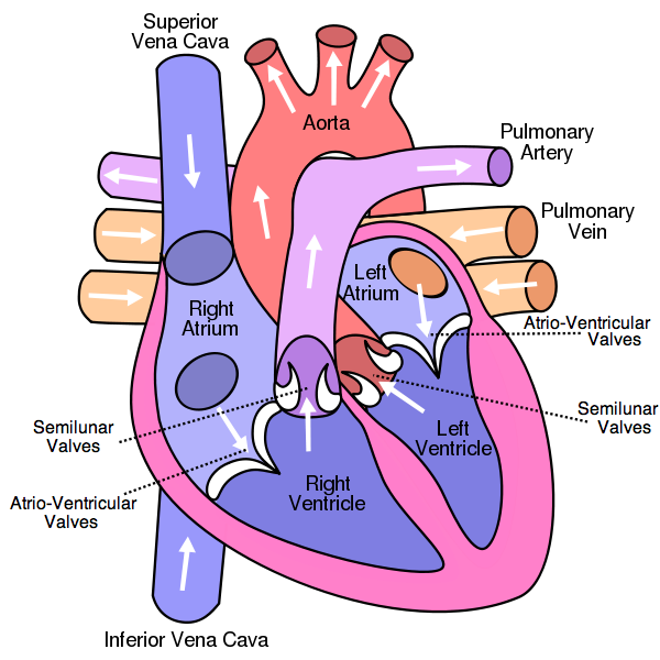
Blood Flow Through The Body Diagram stock illustrations Heart blood flow anatomical diagram with atrium and ventricle system. Vector illustration labeled medical poster. Blood circulation path scheme with arrows. internal organs and circulatory system Vector isolated illustration of human internal organs and circulatory system in obese man body. Stomach, liver, bladder, lung, kidney, heart, icon.

Heart diagram with labels and blood flow
Heart Diagram with Labels and Detailed Explanation - BYJUS The diagram of heart is beneficial for Class 10 and 12 and is frequently asked in the examinations. A detailed explanation of the heart along with a well-labelled diagram is given for reference. Well-Labelled Diagram of Heart The heart is made up of four chambers: The upper two chambers of the heart are called auricles. File:Heart diagram blood flow en.svg - Wikipedia File:Heart diagram blood flow en.svg. Size of this PNG preview of this SVG file: 330 × 370 pixels. Other resolutions: 214 × 240 pixels | 428 × 480 pixels | 535 × 600 pixels | 685 × 768 pixels | 913 × 1,024 pixels | 1,827 × 2,048 pixels. This is a file from the Wikimedia Commons. Information from its description page there is shown below. The Heart | Circulatory Anatomy - Visible Body The Atria Are the Heart's Entryways for Blood The left atrium and right atrium are the two upper chambers of the heart. The left atrium receives oxygenated blood from the lungs. The right atrium receives deoxygenated blood returning from other parts of the body. Valves connect the atria to the ventricles, the lower chambers.
Heart diagram with labels and blood flow. Heart blood flow circulation anatomical diagram with ... - VectorMine Description: Heart blood flow anatomical diagram with atrium and ventricle system. Vector illustration labeled medical poster. Blood circulation path scheme with arrows. You may also like… Coronary circulation anatomical cross section diagram, labeled vector illustration scheme € 7.99 Add to cart Heart Diagram - 15+ Free Printable Word, Excel, EPS, PSD Template ... This type of heart diagram template is a physical representation of a human heart with all its parts mentioned. You can have a high quality picture of this on downloading. 1910 Human Heart Anatomy Print This is an illustration from an old medical book which not only shows the heart diagram but also the concerned blood vessels. The 8 Best Supplements To Boost Blood Flow Naturally - UMZU Dec 04, 2020 · Pulmonary circulation begins when the blood finally reaches the heart. The heart pumps this deoxygenated blood into the lungs through the pulmonary artery. The Red blood cells then take up Oxygen and release CO2. This blood gets returned to the heart through the pulmonary vein, and is then pumped through the systemic circulatory system. YR 8 Topic 2 Circulatory System - Pinterest Diagram of Coronary heart Blood Movement for Cardiac Nursing College students. shoppingdealer. ... The areas of the heart with LESS oxygen are labeled with a "B". Students will color these areas BLUE. ... The Human Circulatory System comprises the heart, blood vessels, blood, lymph, arteries, capillaries and veins. ...
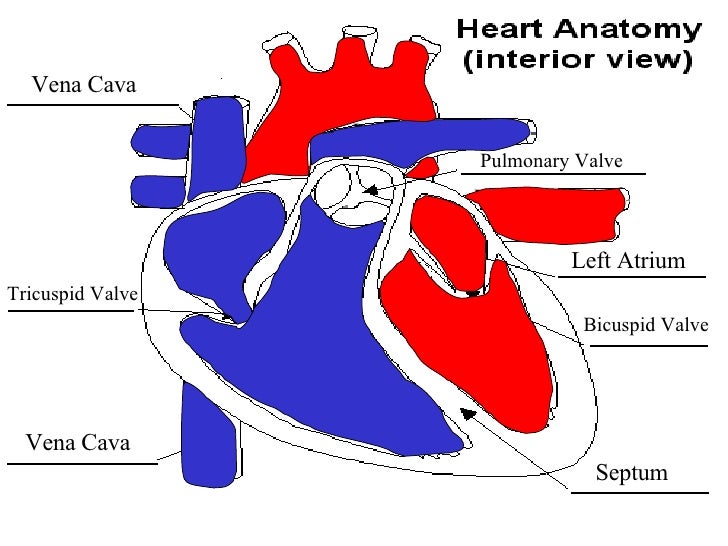
Diagram of Human Heart and Blood Circulation in It Every heart diagram labeledwill clearly show these valves. These valves allow blood flow in one direction only. Different valves perform different functions. Tricuspid valve is located between the right ventricle of your heart and the right atrium, and allows the blood to move from the right atrium to the right ventricle. heart diagram blood flow heart diagram blood flow the Labeled Diagram Of The Heart And Blood Flow blood coming out of we have 9 Pics about the Labeled Diagram Of The Heart And Blood Flow blood coming out of like the Labeled Diagram Of The Heart And Blood Flow blood coming out of, Pin on College/ Nursing and also Pulmonary and Systemic Circulation - YouTube. Here you go: Label the heart — Science Learning Hub Jun 16, 2017 · Labels. Description. Vena cava. Carries deoxygenated blood from the body to the heart. Semilunar valve. Flaps that prevent backflow of blood. Left atrium. Receives oxygenated blood from the lungs. Left ventricle. Region of the heart that pumps oxygenated blood to the body. Pulmonary artery. Carries deoxygenated blood to the lungs. Right ventricle Heart Diagrams for Labeling and Coloring, With ... - Teachers Pay Teachers - One black and white heart diagram with lines for students to fill in labels, and arrows showing blood flow - One black and white heart diagram with no lines or labels, but arrows included, so you can customize what labels the diagram will include - One black and white heart diagram with no lines, labels, or arrows, but with texture of the ...
Diagram Of Body Organs Female Pics Pictures, Images ... - iStock Endometrial polyp or uterine polyp Endometrial polyp or uterine polyp. Sessile polyp and pedunculated polyp. The polyps consist of dense, fibrous tissue, blood vessels and endometrial epithelium. They are attached by a thin stalk or sessile. Vector diagram. diagram of body organs female pics stock illustrations Dopamine - Wikipedia Its effects, depending on dosage, include an increase in sodium excretion by the kidneys, an increase in urine output, an increase in heart rate, and an increase in blood pressure. At low doses it acts through the sympathetic nervous system to increase heart muscle contraction force and heart rate, thereby increasing cardiac output and blood ... Circulatory System Diagram - New Health Advisor This circuit typically includes the movement of blood inside heart and 'myocardium' (the membrane of heart). Coronary circuit mainly consists of cardiac veins including anterior cardiac vein, small vein, middle vein and great (large) cardiac vein. There are different types of circulatory system diagrams; some have labels while others don't. PDF BLOOD FLOW THROUGH THE HEART diagram deoxygenated blood from body tissue oxygenated blood from lungs via pulmonary vein s superior and inferior vena cava left atrium right atrium bicuspid valve tricuspid valve left ventricle ... blood flow through the heart diagram author: taustin created date: 9/30/2013 9:09:13 pm
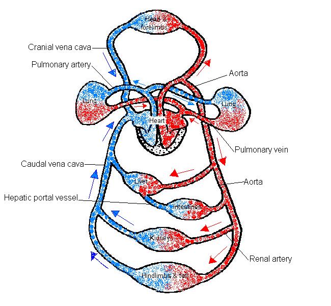
The Anatomy and Physiology of Animals/Circulatory System Worksheet/Worksheet Answers - WikiEducator
Human Heart - Anatomy, Functions and Facts about Heart The external structure of the heart has many blood vessels that form a network, with other major vessels emerging from within the structure. The blood vessels typically comprise the following: Veins supply deoxygenated blood to the heart via inferior and superior vena cava, and it eventually drains into the right atrium.
Free Unlabeled Heart Diagram, Download Free Clip Art, Free Clip Art on Clipart Library
Box Diagram, Labels of Heart, and Blood Flow through Heart About Press Copyright Contact us Creators Advertise Developers Terms Privacy Policy & Safety How YouTube works Test new features Press Copyright Contact us Creators ...
Heart Anatomy and Blood Flow (Advanced)
Heart Anatomy: Labeled Diagram, Structures, Blood Flow ... - EZmed There are 4 chambers, labeled 1-4 on the diagram below. To help simplify things, we can convert the heart into a square. We will then divide that square into 4 different boxes which will represent the 4 chambers of the heart. The boxes are numbered to correlate with the labeled chambers on the cartoon diagram.

mechanism learn and de Labeled Diagram Of The Heart And Blood Flow mai drag and drop the text ...
blood circulation in heart blood circulation in heart. Severe bleeding - The Emergency-Free Home - blood, body, high we have 9 Pics about Severe bleeding - The Emergency-Free Home - blood, body, high like the Labeled Diagram Of The Heart And Blood Flow blood coming out of, Human circulatory system, animation. The heart and arteries are shown and also Transvenous ...
IB Biology Notes - 6.2 The transport system
Blood Throught the Heart - Austin Community College District Blood Flow Through the Heart. Beginning with the superior and inferior vena cavae and the coronary sinus, the flowchart below summarizes the flow of blood through the heart, including all arteries, veins, and valves that are passed along the way. 1. Superior and inferior vena cavae and the coronary sinus. 2.

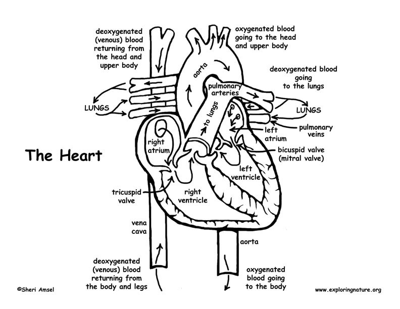
How the Heart Works: Human Heart Diagram, Blood Flow to the Heart & Lung
Practical notes - SP 2.3c Dissection of a Mammalian Heart ... 3. Use a glass rod to follow the path of blood flow : via the pulmonary vein, left atrium and through the bicuspid valve into the left ventricle; via the left ventricle through the semilunar valve and out of the aorta. 4. Note the muscular surface of the ventricle chambers which ensur es smooth blood flow. 5.

Anatomy Of The Heart And Blood Flow - Anatomy Drawing Diagram
A Diagram of the Heart and Its Functioning Explained in Detail The heart blood flow diagram (flowchart) given below will help you to understand the pathway of blood through the heart.Initial five points denotes impure or deoxygenated blood and the last five points denotes pure or oxygenated blood. 1.Different Parts of the Body ↓ 2.Major Veins ↓ 3.Right Atrium ↓ 4.Right Ventricle ↓ 5.Pulmonary Artery ↓ 6.Lungs
Free Blank Heart Diagram, Download Free Blank Heart Diagram png images, Free ClipArts on Clipart ...
Human Heart Diagram Pictures, Images and Stock Photos Heart blood flow anatomical diagram with atrium and ventricle system. Vector illustration labeled medical poster. Blood circulation path scheme with arrows. XXXL Very Detailed Human Heart Engraving From 1872 Featuring A Human Heart. the human heart, the study of anatomy, the path of blood flow in
.png)
Cuthbert - 7th Grade Science Day to Day: Wednesday April 24th - Heart and Blood Flow
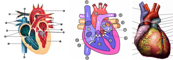
Heart Anatomy & Circulatory System Blood Flow - Human Physiology Two labeled diagrams presenting the anatomy of the heart and the circulatory systems. The top right provides an in-depth diagram of the anatomy of the chambers, valves, and vasculature connected to the heart. The bottom left emphasizes the anatomy of the systemic and pulmonary circulations.
Blood vessels diagram
Diagram of Blood Flow Through the Heart - Bodytomy The heart is divided into two chambers, left and right, the right atrium and ventricle lie on the right side and the left atrium and ventricle on the left side. These two chambers are not directly connected to each other. Synchronization of the Two Chamber The right and left side or chambers of the heart work in tandem with each other.
Blood Flow Animation
Heart Blood Flow | Simple Anatomy Diagram, Cardiac Circulation Pathway ... Step 1 and 6 involve a blood vessel, which makes sense as this is how blood enters and exits that side of the heart. Steps 2-5 involve a chamber, valve, chamber, and valve. So if you remember this general pattern, it will help you recall the order in which blood flows through each side of the heart. Right Side of the Heart SVC/IVC Right Atrium
Blood circulation clipart 20 free Cliparts | Download images on Clipground 2021
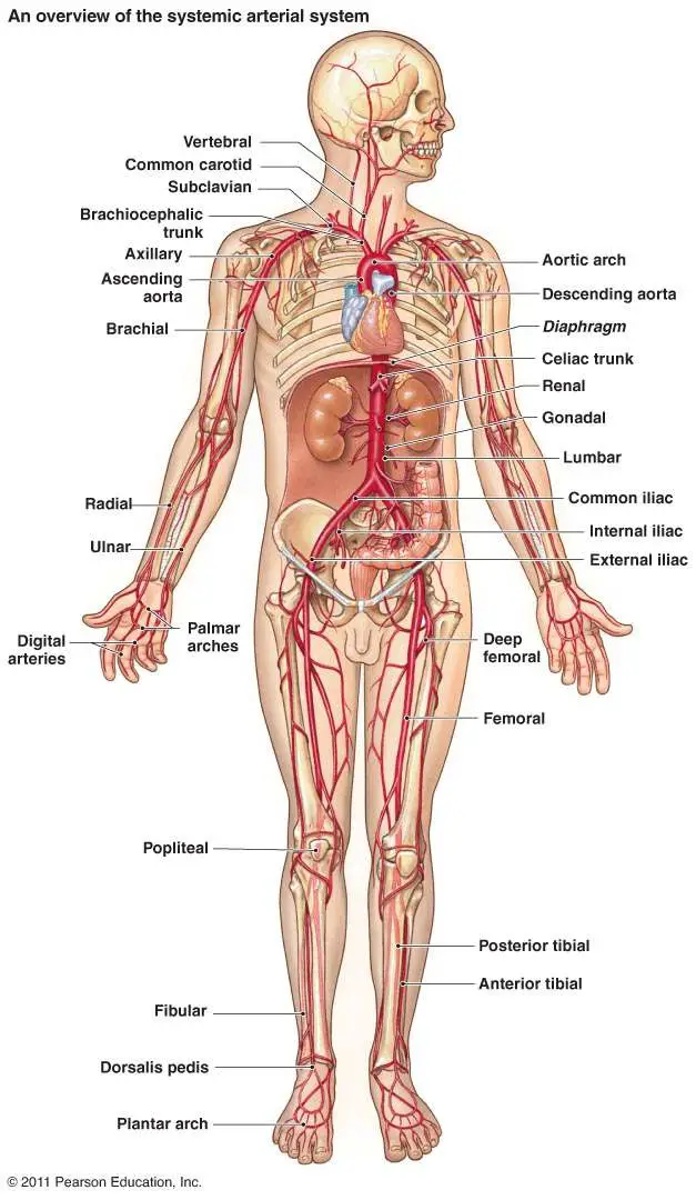
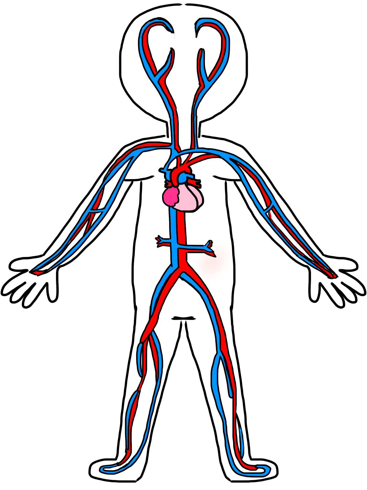
Diagram of Blood Flow in the Heart, Lungs and Body This medical illustration depicts a diagram of blood flow through the body. Oxygenated (oxygen-rich)blood travels from the lungs to the heart, where it is then pumped throughout the body. Deoxygenated (oxygen-poor) blood travels from the body back to the heart, where it is pumped to the lungs for gas exchange. Labels include the common carotid arteries, jugular veins, superior vena cava ...
IB DP Biology Topic 6: Human physiology : 6.2 The blood system Question Bank SL Paper 1
Circulatory System Diagram - Cardiovascular System and Blood ... SmartDraw has a number of templates included for circulatory system diagrams, cardiovascular system diagrams, blood circulation diagrams, and more. You don't really have to "draw" them as much as find them and modify them as needed. You can add labels or titles and change the size of symbols as necessary.
Post a Comment for "40 heart diagram with labels and blood flow"