38 diagram of a human cell with labels
Anatomy (Human Body) Labeling - Exploring Nature Muscles of the Leg and Foot Labeling Page. Muscles of the Neck, Chest and Thorax Labeling Page. Muscles of the Neck, Shoulders and Thorax (Posterior) Labeling. Muscles of the Posterior Body Labeling (HS-Adult) Muscles of the Thigh and Hip (Anterior) Labeling. Muscles of the Thigh and Hip (Posterior) Labeling. Nerve Cell (Neuron) Labeling Page. Cell: Structure and Functions (With Diagram) - Biology Discussion Eukaryotic Cells: 1. Eukaryotes are sophisticated cells with a well defined nucleus and cell organelles. 2. The cells are comparatively larger in size (10-100 μm). 3. Unicellular to multicellular in nature and evolved ~1 billion years ago. 4. The cell membrane is semipermeable and flexible. 5. These cells reproduce both asexually and sexually.
Label Cell Parts | Plant & Animal Cell Activity | StoryboardThat Create a cell diagram with each part of plant and animal cells labeled. Include descriptions of what each organelle does. Click "Start Assignment". Find diagrams of a plant and an animal cell in the Science tab. Using arrows and Textables, label each part of the cell and describe its function.

Diagram of a human cell with labels
Human Cell Organelles Labeling Diagram - Quizlet Human Cell Organelles Labeling STUDY Learn Flashcards Write Spell Test PLAY Match Gravity Created by Mackenna_Rios5 Terms in this set (8) Vesicles Transports molecules between organelles and the cell membrane Ribosome Makes Protein Mitochondria Makes ATP Smooth ER Makes lipids and vesicles Lysosomes PDF Human Cell Diagram, Parts, Pictures, Structure and Functions Diagram of the human cell illustrating the different parts of the cell. Cell Membrane The cell membraneis the outer coating of the cell and contains the cytoplasm, substances within it and the organelle. It is a double-layered membrane composed of proteins and lipids. Animal Cell Diagram with Label and Explanation: Cell Structure, Functions Below is the diagram of the animal cell which shows the organelles present in it. The cell is covered with cytoplasm which consists of cell organelles in it. The nucleus is covered with a rough Endoplasmic Reticulum and other organelles each designed for a specific purpose.
Diagram of a human cell with labels. Cell Membrane Diagram Labeled : Functions and Diagram Cell Membrane Diagram. There are no organelles in the prokaryotic cells, i.e., they have no internal membrane systems. While lipids help to give membranes their flexibility, proteins monitor and maintain. We all keep in mind that the human body is very elaborate and a technique I found out to understand it is by means of the manner of human ... Labeled Diagram of the Human Kidney - Bodytomy Labeled Diagram of the Human Kidney The human kidneys house millions of tiny filtration units called nephrons, which enable our body to retain the vital nutrients, and excrete the unwanted or excess molecules as well as metabolic wastes from the body. In addition, they also play an important role in maintaining the water balance of our body. Animal Cells: Labelled Diagram, Definitions, and Structure Animal Cells Organelles and Functions. A double layer that supports and protects the cell. Allows materials in and out. The control center of the cell. Nucleus contains majority of cell's the DNA. Popularly known as the "Powerhouse". Breaks down food to produce energy in the form of ATP. Human Cell Diagram, Parts, Pictures, Structure and Functions - Pinterest This diagram of a human skeleton is labeled with 12 major bones, from skull to fibula. D Nicole Science Plant Cell Drawing Animal Cell Drawing Human Cell Diagram Plant Cell Diagram Biology Drawing Science Drawing Biology Art Human Drawing Science Biology Plant cells are covered by cell wall, it is a unique feature observed in plant cells.
117 Skin Cell Diagram Premium High Res Photos - Getty Images Browse 117 skin cell diagram stock photos and images available, or start a new search to explore more stock photos and images. Psoriasis in section, skin with detailed epidermis in stratum corneum, granular and germinative, dermis, and hypodermis. Labeled Diagram Of The Skeletal System Pic Stock Photos, Pictures ... Labeled Anatomy Chart of Shoulder, Elbow and Triceps Muscles in... Labeled human anatomy diagram of man's shoulder bones, triceps muscles and connective tissue in a posterior view on a black background. Diagram of a longitudinal section of a long bone Illustration of a Diagram of a longitudinal section of a long bone Development of a long bone A Labeled Diagram of the Plant Cell and Functions of its Organelles Unique to plant cells, the cell wall is a fairly rigid, protective wall that resists the strain of physical forces. The cell wall is mainly made up of cellulose fiber and it helps maintain the shape of the cell. Function: Maintains cell pressure and prevents over-expansion of the cell. Centrosome What Is Going On Inside That Cell? | Human cell diagram, Cell diagram ... Source: Shutterstock Technology has brought advances that were previously unheard of just a couple of decades ago. One of which is the ability to look inside of a cell. Yes, a cell. Think of your biology class with the posters or charts on the wall with life size versions of the inner components of the cell which reside at distances on the ...
File:Diagram human cell nucleus tr.svg - Wikimedia Commons Description. Diagram human cell nucleus tr.svg. en: A diagram of a human cell nucleus, with Turkish labels. Translated version of File:Diagram human cell nucleus.svg, originally created and all rights released by Mariana Ruiz ( User:LadyofHats ). This image is also released to the public domain. az: İnsan hüceyrə nüvəsinin sxematik rəsmi ... Cells Diagram | Science Illustration Solutions - Edrawsoft Cells Diagram. Cells are the basic building blocks of all living things. The human body is composed of trillions of cells. Cells have many parts, each with a different function. Some of these parts, called organelles, are specialized structures that perform certain tasks within the cell. Drawing cells diagram helps you better understand your ... Human Cells Printables and Diagrams - The Successful Homeschool These cells include: leukocytes, haematids, thrombocytes, ovum, sperm, sarcomeres, enterocytes, neurons, osteocytes, hepatocytes. They will learn the parts of a cell thanks to a labeled diagram. They will get to see what blood looks like under a microscope without needing to own a microscope. They get to color a cell and then label the parts. Animal Cell Diagram | Science Trends An animal cell diagram is a great way to learn and understand the many functions of an animal cell. The diagram, like the one above, will include labels of the major parts of an animal cell including the cell membrane, nucleus, ribosomes, mitochondria, vesicles, and cytosol.
Free vector graphic: View, Cell, Information, Close - Free Image on Pixabay - 48543
The Human Skeleton: All You Need to Know - Bodytomy Labeled Skeleton Diagram This skeleton diagram will help explain the different bones of the human body clearly. Cranium The cranium is a skull bone that covers the brain, as seen in the skeleton diagram. The facial bones are not a part of the cranium. The bones that are just above the ear or in front of the ear are known as temporal bones. Stapes

Follow me: Arts
Label Diagram Human Body Stock Illustrations - Dreamstime Download 161 Label Diagram Human Body Stock Illustrations, Vectors & Clipart for FREE or amazingly low rates! New users enjoy 60% OFF. 185,925,055 stock photos online. ... Animal cell structure anatomy infographic diagram. With parts flat vector illustration design for biology science education school book concept microbiology.

a. Cell : unit of function - BIOLOGY4ISC
Learn the parts of a cell with diagrams and cell quizzes - Kenhub For this exercise we'll start with an image of a cell diagram ready labeled. Study this and make sure that you're clear about which structure is found where. Cell diagram unlabeled It's time to label the cell yourself! As you fill in the cell structure worksheet, remember the functions of each part of the cell that you learned in the video.

what is ureter 744×1240 | Anatomy System - Human Body Anatomy diagram and chart images
Human Cell Diagram, Parts, Pictures, Structure and Functions One of the few cells in the human body that lacks almost all organelles are the red blood cells. The main organelles are as follows : cell membrane endoplasmic reticulum Golgi apparatus lysosomes mitochondria nucleus perioxisomes microfilaments and microtubules Diagram of the human cell illustrating the different parts of the cell. Cell Membrane

Anatomy of a human cell with labels for individual organelles | Organelles, Medical illustration ...
Human Heart Diagram Labeled | Science Trends Daniel NelsonPRO INVESTOR. The human heart is an organ responsible for pumping blood through the body, moving the blood (which carries valuable oxygen) to all the tissues in the body. Without the heart, the tissues couldn't get the oxygen they need and would die. Along with lymphatic vessels, the blood, blood vessels, and lymph, the heart ...

cell labeled | HUMAN ANATOMY & PHYSIOLOGY | Pinterest
Diagram of human skin structure - Science Learning Hub Diagram of human skin structure. Add to collection. + Create new collection. Tweet. Rights: University of Waikato Published 1 February 2011 Size: 100 KB Referencing Hub media. The epidermis is a tough coating formed from overlapping layers of dead skin cells.

Human Behavior and the Social Environment I BSW: CELL DIAGRAM
Human Cell Coloring Page | crayola.com Human Cell. Use Crayola® crayons, colored pencils, or markers to color the parts of the cell. Use the word bank below to identify the parts of the human cell.The cell is the basic unit of the human body. There are over one billion cells in each human body. Cells group together to make skin, bones, and blood.
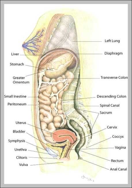
uterus location | Anatomy System - Human Body Anatomy diagram and chart images
File:Diagram human cell nucleus.svg - Wikipedia A comprehensive diagram of a human cell nucleus. Date: 27 April 2006: Source: I did it myself with adobe ilustrator using the information found here , ,, and : Author: Mariana Ruiz LadyofHats: Permission (Reusing this file) Public domain Public domain false false:

Xtra! Xtra! Read All About It Newspaper!: Biology: Learning About Cells
Circulatory System Labeled Diagram stock illustrations Browse 154 circulatory system labeled diagram stock illustrations and vector graphics available royalty-free, or start a new search to explore more great stock images and vector art. Newest results Heart Poster Heart blood flow circulation anatomical diagram with atrium and... Anatomy of Nerves of Body and Head
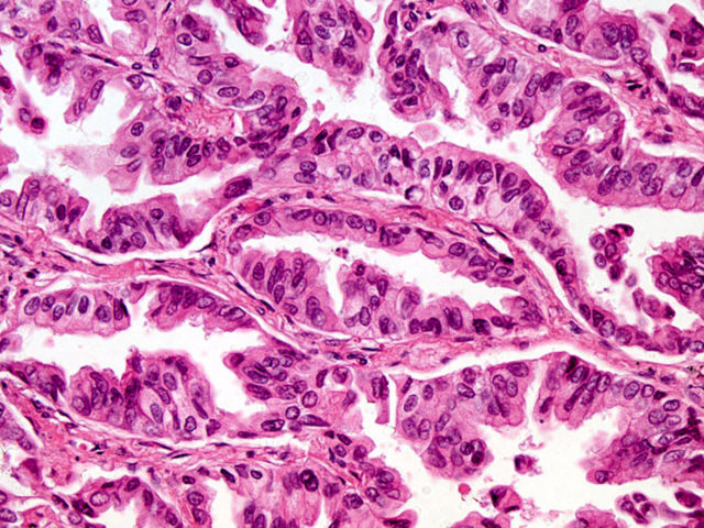
Human Pathology | Nikon’s MicroscopyU
A Labeled Diagram of the Animal Cell and its Organelles As observed in the labeled animal cell diagram, the cell membrane forms the confining factor of the cell, that is it envelopes the cell constituents together and gives the cell its shape, form, and existence. ... it is essential that the DNA remains intact and gets evenly distributed among the cells. Every human body cell contains 46 ...

Luke's Place | This blog is about my school year and myself.
Animal Cell Diagram with Label and Explanation: Cell Structure, Functions Below is the diagram of the animal cell which shows the organelles present in it. The cell is covered with cytoplasm which consists of cell organelles in it. The nucleus is covered with a rough Endoplasmic Reticulum and other organelles each designed for a specific purpose.

Science
PDF Human Cell Diagram, Parts, Pictures, Structure and Functions Diagram of the human cell illustrating the different parts of the cell. Cell Membrane The cell membraneis the outer coating of the cell and contains the cytoplasm, substances within it and the organelle. It is a double-layered membrane composed of proteins and lipids.

Human Cell Diagram To Label - General Wiring Diagram
Human Cell Organelles Labeling Diagram - Quizlet Human Cell Organelles Labeling STUDY Learn Flashcards Write Spell Test PLAY Match Gravity Created by Mackenna_Rios5 Terms in this set (8) Vesicles Transports molecules between organelles and the cell membrane Ribosome Makes Protein Mitochondria Makes ATP Smooth ER Makes lipids and vesicles Lysosomes
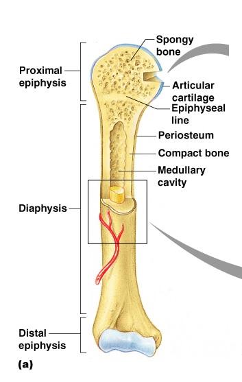
Print Exercise 9: Overview of the Skeleton: Classification and Structure of Bones and Cartilages ...
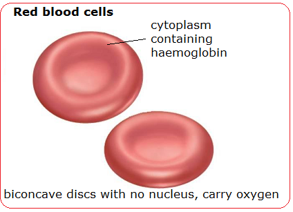
Blood cells - structure and functions - Biology Notes for IGCSE 2014
PRACTICAL BOOKLET - BIOLOGY4ISC

veins and arteries of the leg | Anatomy System - Human Body Anatomy diagram and chart images
Post a Comment for "38 diagram of a human cell with labels"